
Heart Restart
Every 40 seconds, someone in the U.S. has a heart attack. While not always fatal, the damage to heart tissue is irreversible. In contrast, zebrafish have the ability to regenerate their heart tissue and return their hearts to a nearly normal state.
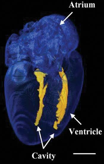
Bioengineering Assistant Professor Juhyun Lee wants to know why. He is using a three-year, $450,000 grant from the National Science Foundation to develop an imaging technique and a special electrocardiogram (EKG) for zebrafish to determine how they are able to regenerate their heart tissue after cardiac arrest.
“Contractility is very important for proper heart function, but scar tissue stiffens the human heart and reduces how much it can contract to pump blood,” Dr. Lee says. “In zebrafish, the scars heal much faster and more completely than other animals, so we’re trying to find out why and how.”
He and his team have developed a special EKG jacket for the fish, in collaboration with researchers at UC Irvine. Once the fish are fitted with the EKG jacket, Lee uses a superresolution ultramicroscope to create 3D images of their hearts. Using a clearing method, PEGASOS, that he developed with colleagues at Texas A&M University, Lee makes the tissue samples transparent to easily see through the inside of the entire heart with the microscope. This way, he can monitor the proliferation of cardiomyocytes to determine the importance of biomechanical force in their development.